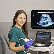
HOW Do Ultrasound Scans Work

Ultrasound scans, also known as sonography, work by using high-frequency sound waves to create images of the inside of the body. Here’s a step-by-step explanation of how ultrasound scans work:
- Generation of Sound Waves: A transducer, a handheld device, is used to generate high-frequency sound waves. These sound waves are typically inaudible to the human ear, with frequencies higher than the upper audible limit of human hearing.
- Transmission into the Body: The transducer is placed against the skin on the area being examined. A gel is often applied to the skin to ensure proper contact and to eliminate air pockets that can interfere with the transmission of sound waves.
- Propagation through Tissues: The generated sound waves travel through the body tissues. When they encounter different types of tissues (fluids, solids, air), some of the waves are reflected back to the transducer while others continue to penetrate deeper.
- Echo Reception: The transducer not only sends out sound waves but also receives the echoes created as the waves bounce back from the internal structures. The timing and intensity of these echoes provide information about the density and composition of the tissues.
- Conversion into Images: The received echoes are converted into electrical signals. These signals are then processed by a computer, which uses the information to create real-time images on a monitor. The images show the internal structures, such as organs or a developing fetus during pregnancy.
- Doppler Ultrasound (optional): In some cases, a Doppler ultrasound may be used to assess blood flow. This involves evaluating the Doppler shift in the frequency of the echoes, allowing visualization of blood movement in vessels.
- Recording or Capturing Images: The images can be recorded or captured in real-time during the examination. This helps healthcare professionals review and analyze the structures and functions of the internal organs or monitor the progress of a pregnancy.
Ultrasound scans are widely used in various medical applications, including monitoring pregnancies, imaging abdominal and pelvic organs, and assessing blood flow in vessels. They are non-invasive, safe, and do not involve ionizing radiation, making them a valuable diagnostic tool in medical practice.
if you are preparing Ultrasound exam. join ultrasound course in StudyUltrasound . StudyUltrasound is the best option.

Comments
Post a Comment