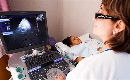
Learn How Different Ultrasound Scans Work and What Their Use Is

A sonogram is another name for an ultrasound scan. During this process, high-frequency sound waves are used to create an image of a portion of the interior of the body.
There are numerous uses for ultrasound. It can be used to guide a surgeon during specific surgeries, diagnose a problem, and keep an eye on an unborn child.
Working Of Ultrasound
An ultrasonic probe, a tiny gadget, is employed. High-frequency sound waves are emitted by this. It is impossible for you to hear these waves of sound. The “echoes” that are produced when sound waves reverberate off various body parts are detected by the probe and converted into a moving picture.
While the scan is running, this image is shown on a monitor.
Different Kinds of Ultrasound Scans
Different types of ultrasound scanning exist. Which body part is being scanned and for what purpose determines the kind in large part.
The three primary categories are:
External Ultrasound Scan
The main purposes of an external ultrasound scanning are to look at an unborn child’s heart or womb. In addition, it can be used to evaluate other organs or tissues that are visible through the skin, like muscles and joints, as well as the liver, kidneys, and other organs located in the abdomen and pelvis.
Your skin is probed on the area of your body that is being checked with a tiny, portable device. Your skin is covered with a lubricating lubricant to facilitate the smooth movement of the probe. This guarantees that the probe and skin remain in constant contact.
Internal or Transvaginal Ultrasound Scan
An internal examination gives a physician a closer look at organs like the womb, ovaries, and prostate gland inside the body.
Meaning “through the vagina,” an ultrasound is called transvaginal. You will be instructed to lie on your side with your knees pulled up towards your chest, or on your back, during the treatment.
Images are then sent to a monitor using a tiny, sterile ultrasonic probe that is little wider than a finger. The probe is then gently inserted into the vagina or rectum.
Internal examinations typically don’t hurt and shouldn’t take too long, but they may cause some discomfort.
Endoscopic Ultrasound Scan
An endoscope is inserted into your body to perform an endoscopic ultrasound scan. In order to examine places like your stomach or food pipe, this is typically done through the mouth (oesophagus).
As the endoscope is gently lowered towards your stomach, you will be requested to lie on your side. The end of the endoscope is equipped with an ultrasonic instrument and a light. Similar to an external ultrasound, sound waves are employed to produce images once it is put into the body.
A local anaesthetic spray to numb your throat and a sedative to keep you calm are frequently administered because endoscopic ultrasound scans can be painful and even make you feel sick.
In the unlikely event that you bite the endoscope, a mouth guard might be provided to you to keep your mouth open and shield your teeth.
Hope you found this information helpful!!if you want to build a career in ultrasound you can join ultrasound courses in StudyUltrasound

Comments
Post a Comment